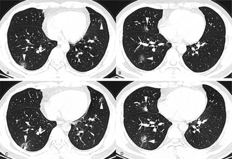
Figure 4.

A-B. Baseline chest CT images of a 35-year-old male with a negative first RT-PCR test, presenting multiple patchy areas of pure GGOs and GGOs with interlobular septal thickening in both lungs. C-D. Two days after baseline chest CT, when he had his first positive RT-PCR test, CT images showed that the consolidation component had increased (white arrowhead).
