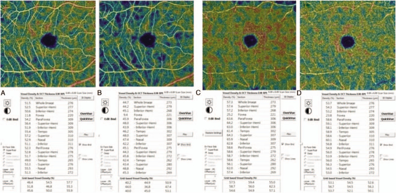
Figure 2.

Images of the vessel densities measured before and one hour after the laser peripheral iridotomy (LPI). A: Measurements of the superficial retinal layer at the baseline; B: Measurements of the superficial retinal layer at one hour after LPI; C: Measurements of the deep retinal layer at the baseline; D: Measurements of the deep retinal layer at 1 h after the LPI.
