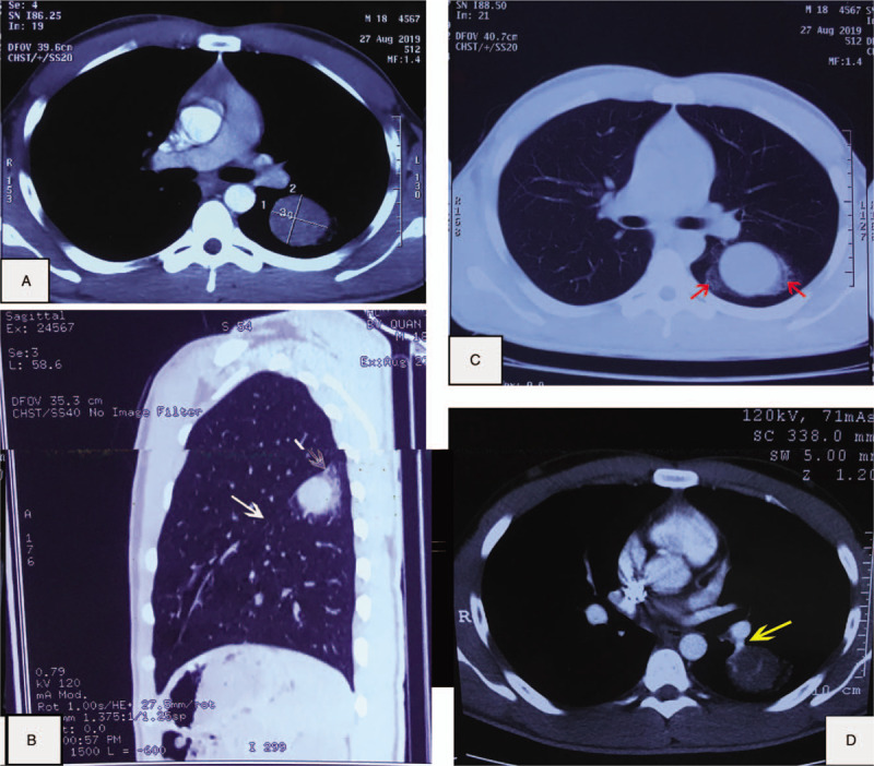
Figure 1.

(A and B) A round lesion located in the lower left lobe in thorax CT, below the left greater fissure (white arrows). (C) A mass with surrounding ground-glass opacity (red arrows), defined as the “halo sign.” (D) CT performed 1 mo later revealed that the size had not changed. An obviously enhanced, engorged vascular structure (a yellow arrow) adjacent to the lesion. It was defined as the “overlying vessel sign.” CT = computed tomography.
