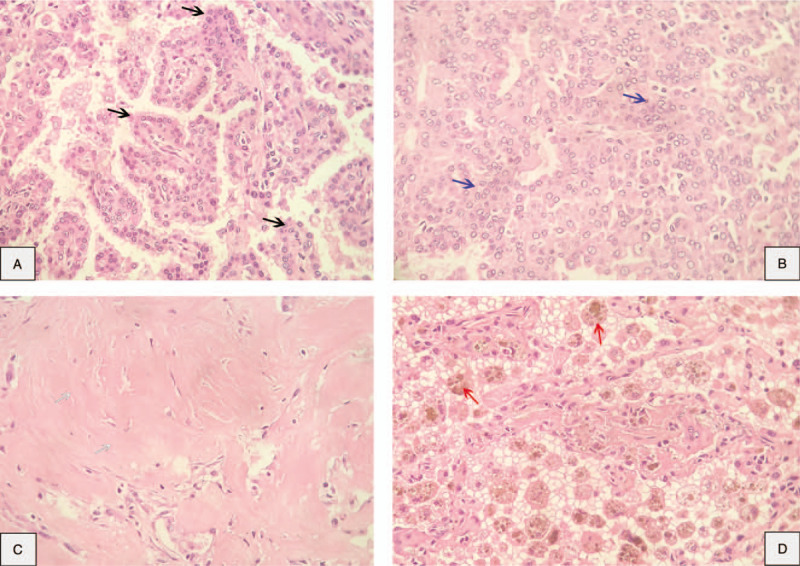
Figure 2.

Pathological findings: pulmonary sclerosing pneumocytoma in a core-needle biopsy (hematoxylin-eosin, 40×). Two types of cells, cuboidal surface cells and stromal round cells, were organized into 4 structural patterns. (A) Papillary (black arrows). (B) Solid (blue arrows). (C) Sclerotic (white arrows). (D) Hemorrhagic (red arrows).
