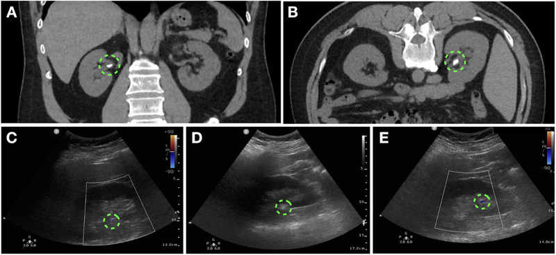
FIGURE 1.

Repositioned ureteropelvic junction stone. (A) Coronal and (B) axial CT images with patient prone demonstrate a 9‐mm right ureteropelvic junction stone with mild hydronephrosis. A coronal ultrasound image prior to stone repositioning (C) shows the same 9‐mm echogenic calcification exhibiting the twinkling artifact at the ureteropelvic junction with no associated hydronephrosis. After hydration, the ultrasound image (D) reveals the same twinkling ureteropelvic junction stone and moderate hydronephrosis as dilation of the hypoechoic region within the kidney. After stone repositioning, (E) shows the stone apparent as echogenic and twinkling now in the lower pole of the kidney. Resolution of hydronephrosis is also seen. Supporting Information Videos S1–S3 correspond to the images (C)–(E)
