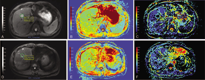
Figure 2.

MR images of a 40 yr-old male patient with colon cancer liver metastases before (upper line) and 1 mo after (lower line) radiofrequency ablation (RFA). Following drawing region-of-interest (ROI) of index tumor and ablative zone on b value of 0 diffusion-weighted imaging (DWI) image (A, D), the apparent diffusion coefficient (ADC) maps (B, E) demonstrated slight increasing of mean ADC value from 1.16 × 10⁻³ mm2/s to 1.22 × 10⁻³ mm2/s. And the f maps (C, F) showed minor increasing of mean f value from 0.21 to 0.28.
