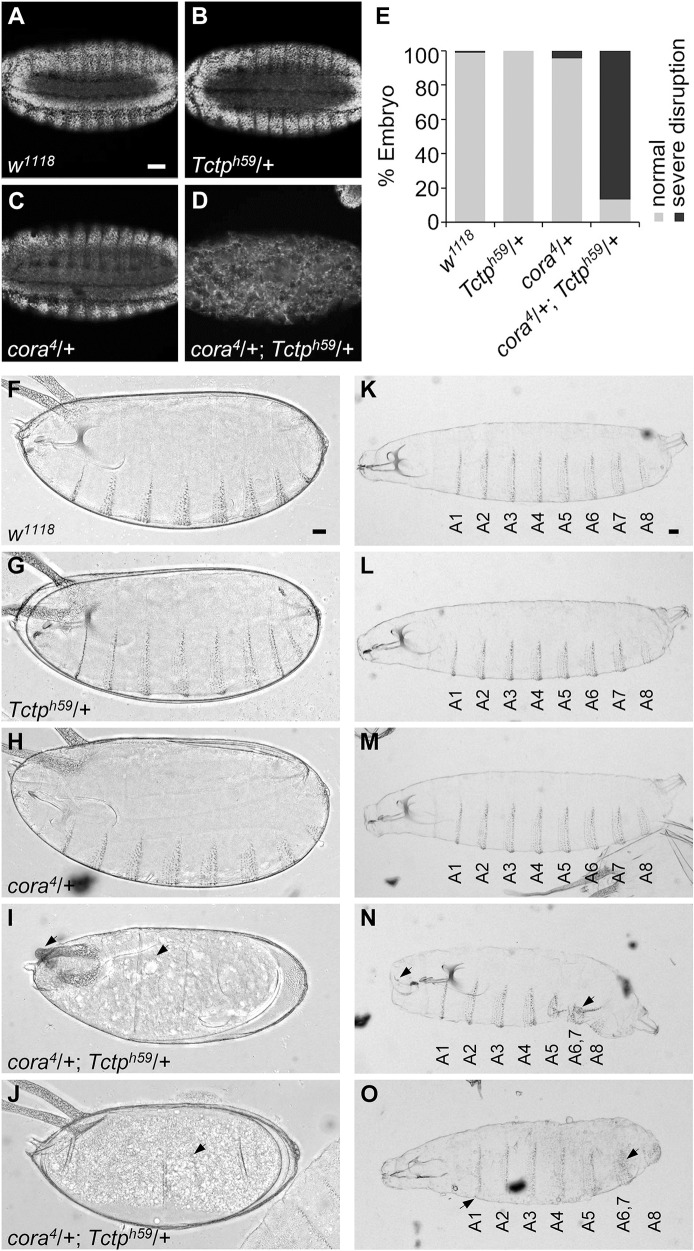
Fig 2. cora and Tctp heterozygotes show synthetic lethality in the embryo.
(A-D) Optical cross-section views of the DAPI staining pattern. Embryo genotypes are as indicated. Tctph59/+ (B) or cora4/+ (C) heterozygotes show relatively normal pattern. Double heterozygous embryos are grossly disorganized (D). (E) Quantification of embryos showing normal morphology and abnormalities. Wild-type and single heterozygous embryos are mostly normal. In contrast, about 87% of double heterozygous embryos show embryo lethality with severe disruption of epithelia. All embryos are at stage 16 (n ≥ 50 for each genotype). (F-O) Denticle phenotypes in late-stage embryos (F-J) and 1st instar larvae (K-O). Wild-type embryo and larva have normal denticles (F, K). Heterozygotes of Tctph59/+ (G, L) or cora4/+ (H, M) show a normal pattern of denticles. cora/+; Tctp/+ double heterozygotes die before denticle formation (I, J, arrows). The only survived larvae show fused or decreased number of denticle belts in the posterior segments (N, O, arrows). Scale bars, 50 μm.

