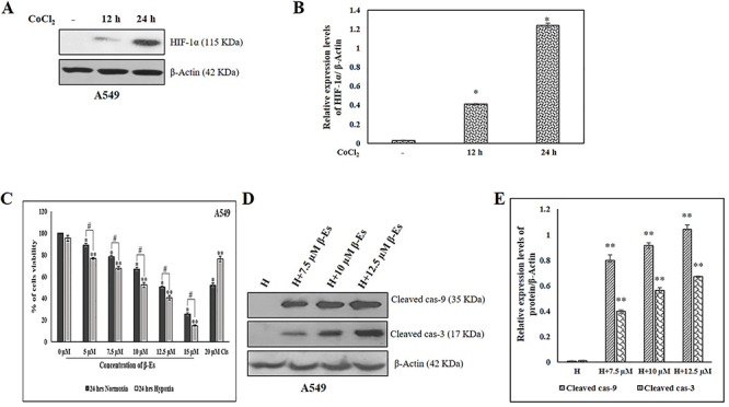
Figure 1.

Effect of β-Es on the viability of A549 cells under normoxia and CoCl2 induced hypoxic condition. (A) A549 cells were incubated under 100 μM CoCl2 for 0, 12 and 24 h. Immunoblot shows HIF-1α expression. (B) Densitometric analysis of HIF-1α protein expression. (C) Effect of β-Es on A549 cells viability under normoxia and 100 μM CoCl2 induced hypoxic condition: A549 cells were prior to exposed the 100 μM CoCl2 for 2 h before treated to various concentration of β-Es treatment for 24 h. Cells viability was determined by MTT assay. (D) A549 cells were pre-incubated 100 μM of CoCl2 for 2 h before treated with β-Es (24 h) for the indicated concentration. At the end of the treatment, cells were lysed and equal amounts of proteins were loaded and western blotting for the detection of cleaved cas-9 and 3. (E) Expression of cleaved cas-9 and 3 were quantified using Image-J software and displayed in bar graph (mean ± SD, n=3). Results were normalized to β-actin. Data are expressed as mean α S.D. *p<0.05 as compared with normoxia; **p<0.05 as compared with hypoxia; #p<0.05 as compared with the β-Es treated normoxia and hypoxia group.
