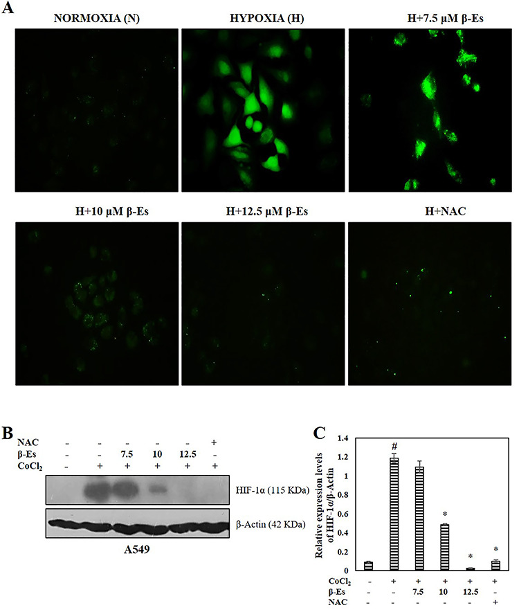
Figure 3.

β-Es repress CoCl2 induced HIF-1a stabilization through the suppression of ROS (A) Fluorescence microscopic images of DCFH-DA stained β-Es treated A549 cells with indicated concentration (20 X magnification). 10?mM NAC used as a positive control. (B) Cell lysates were subjected to western blot analysis with HIF-1α antibodies. β-actin was used as the loading control. (C) A densitometric analysis was used to estimate the expression levels of HIF-1α. Significance: #p<0.05 vs normoxia; *p<0.05 vs hypoxia.
