Figure 7.

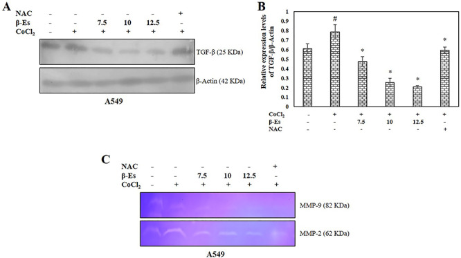
β-Es suppresses TGF-β expression and MMPs activation under CoCl2 induced hypoxic condition (A) Whole cell lysates of β-Es treated A549 cells were immunoblotted with antibodies for TGF-β under conditions of normoxia (N) or hypoxia (H) for 24 h. (B) The mean ± SD of values obtained from densitometric analysis of TGF-β (n=3). Significance: #p<0.05 vs normoxia; *p<0.05 vs hypoxia. (C) The MMP-2 and 9 activities: Cells were pretreated with or without 100 μM CoCl2 for 2 h before treated to β-Es for 24 h. The conditioned media were collected and analyzed by gelatin zymography.
