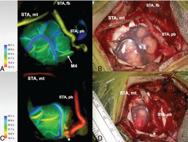
FIGURE 1.

Indocyanine green (ICG) angiography color coded flow 800 (A) and microscopic image (B) before STA-MCA bypass after harvest of the STA, craniotomy and dura opening. The marked M4 MCA branch was selected for the bypass as recipient. STA, superficial temporal artery, STA mt, STA main trunk, STA fb, STA frontal branch, STA pb, STA parietal branch. ICG angiography color coded flow 800 (C) and microscopic image (D) after STA-MCA bypass with patent anastomosis and increased flow compared to pre-bypass.
