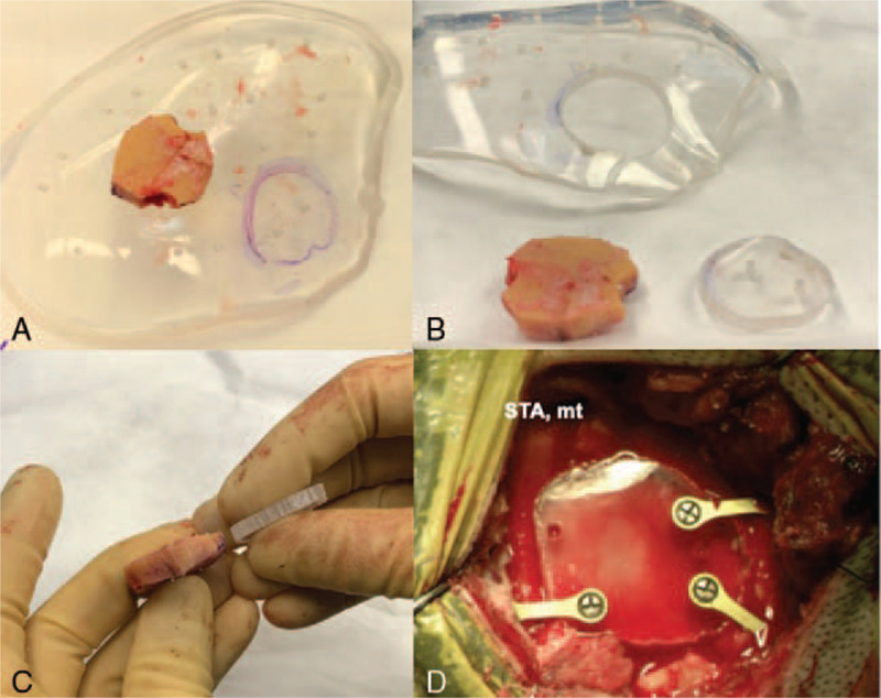
FIGURE 2.

The bone flap was used to determine the size and shape of the clear PMMA implant (A) and an approximately 3 × 3 cm cranioplasty implant was cut out (B, C) and fixated over the craniotomy defect using titanium screws and mini-plates allowing enough space inferiorly without compression of the STA graft moving from extra- to intracranial. STA mt, superficial temporal artery main trunk.
