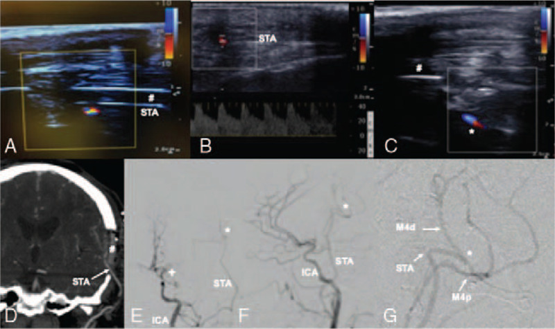
FIGURE 3.

Transcranioplasty ultrasound confirmed flow in donor STA under the cranioplasty (#) in real-time (A), allowed for quantitative flow measurements of the proximal STA donor vessel (B) and confirmed patency of the anastomosis (C). Postoperative CTA coronal view (D) and catheter angiogram in PA (E) lateral (F-G) planes confirmed bypass patency. ∗ bypass anastomosis, + moyamoya disease vessel occlusion. # transparent PMMA cranioplasty implant.
