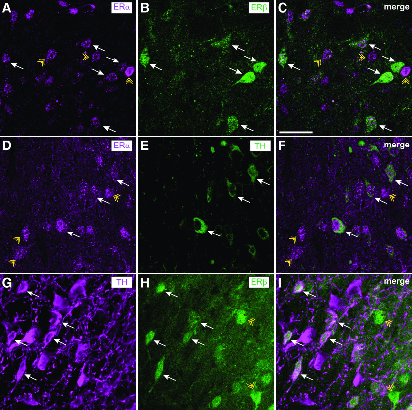
Figure 1.
ERα and ERβ are expressed in DA neurons and nondopaminergic cells in the VTA. Brain sections containing the VTA from female mice in estrus were processed for fluorescent IHC with antibodies to ERα, GFP (reporter for ERβ expression), and TH. A-C, Representative images showing ERα (magenta) and GFP (green) colocalization in the VTA of an ERβ-GFP reporter mouse. White arrows indicate examples of ERα+/GFP+ cells. Yellow arrowheads indicate examples of ERα+/GFP– cells. D-F, Representative images showing ERα (magenta) and TH (green) colocalization in the VTA of a C57BL/6J mouse. White arrows indicate examples of ERα+/TH+ cells. Yellow arrowheads indicate examples of ERα+/TH– cells. G–I, Representative images of TH (magenta) and GFP (green) colocalization in the VTA of an ERβ-GFP reporter mouse. White arrows indicate examples of TH+/GFP+ cells. Yellow arrows indicate examples of TH–/GFP+ cells. Scale bar, 50 μm.

