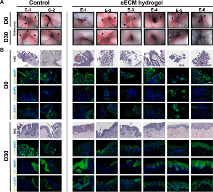
Fig. 4. Effect of eECM hydrogel treatment on the macroscopic appearance of the mucosa and esophageal epithelial cell phenotype after 30 days.

(A) Endoscopies of the lower esophagus/GEJ taken at D0 (before treatment) and at D30 (after treatment) for control and eECM hydrogel animals. Dogs had varied degrees of esophagitis and ulceration (arrowheads) before treatment. No improvement was seen in the animals that only received omeprazole (control). Improvement was seen in all animals after 30 days of eECM hydrogel treatment, including some with complete macroscopic resolution (E-1, E-3, E-4, and E-6). For the remaining treatment animals (E-2 and E-5), there was partial or total resolution of esophagitis with partial healing of the biopsy site. Photo credit: Juan Diego Naranjo, University of Pittsburgh. (B) Biopsies were taken before treatment (D0), and location-matched samples were collected after 30 days of treatment (D30) for control animals and eECM hydrogel–treated animals. Samples at D0 and D30 were stained with hematoxylin and eosin (H&E), Barrett’s marker Sox9, or normal esophageal squamous epithelial markers CK13 and CK14. Arrows indicate goblet cells characteristic of intestinal metaplasia and BE. Scale bar, 100 μm.
