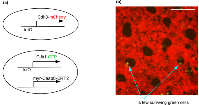
Figure 5. Patterning followed by morphogenesis.
(a) shows the genetic constructs in the two cell types. The cells generated a phase separation pattern on induction of the adhesion systems with tetracycline, to inhibit the TetR repressor protein that is constitutively expressed in these cells. Then, when tamoxifen is added to the medium, the Caspase8-ERT2 induces apoptosis in the Cdh1, green cells, to leave holes in a sieve-like ‘tissue’. Data are from our image sets for [25]; scale bar 200 µm.

