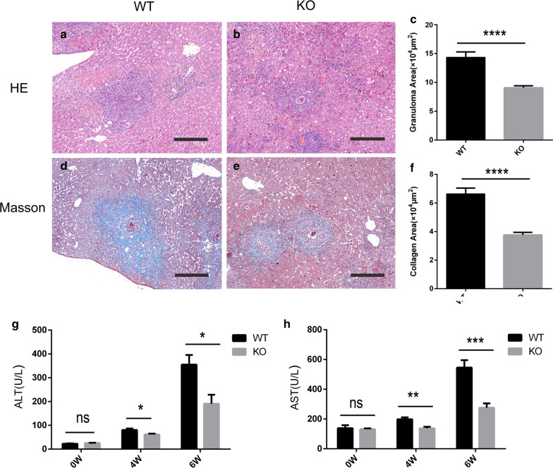
Fig. 3.
Decreased fibrosis and liver damage in S. japonicum-infected TCR δ KO mice. a, b H&E staining showing liver granuloma in WT (a) and TCR δ KO (b) mice at 6 weeks post-infection. c Statistical analysis of the granuloma area between the two groups. d, e Masson staining showing the liver fibrosis in WT (d) and KO (e) mice at 6 weeks post-infection. f Statistical analysis of the fibrosis area between the two groups. g, h Statistical analysis of serum ALT (g) and AST (h) in WT and KO mice at 0, 4 and 6 weeks post-infection. Differences were analyzed by using a Student’s t-test, n = 5 per group *P < 0.05, **P < 0.01, ***P < 0.001, ****P < 0.0001; ns, no significant difference

