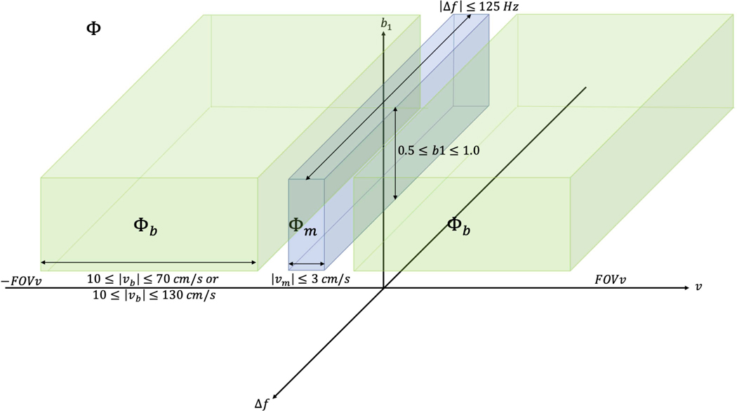
Figure 1:
Cartoon representation of , which includes velocities over ±FOVv, off-resonance values across ±125 Hz, and RF transmit scaling from 1–0.5. , where includes only low-velocities anticipated over the myocardium during stable diastole. , where includes the range of expected coronary arterial blood velocities . represents the velocity vector component encoded along the z-direction. While this whole space is considered for optimization, later figures only show ASL signal as a function of off-resonance for b1=1, and ASL signal as a function of b1 scale for .

