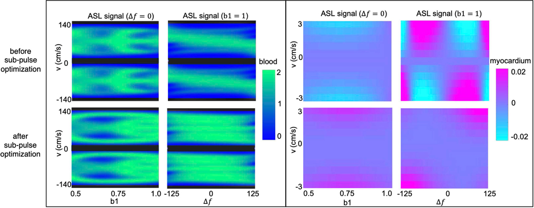
Figure 3:
Bloch simulations of the proposed VS pulse with FOVv =140 cm/s for coronary (left) and for myocardial velocities before (top) and after (bottom) optimization of sub-pulse amplitudes. Bloch simulations are shown for and as well as and . The VS pulse achieves better labeling efficiency of flowing signal (green regions) after optimization, especially for different off-resonance values. In addition, it can be seen that the VS pulse allows for less spurious labeling of moving myocardium (purple regions) after optimization.

