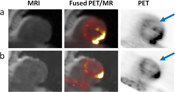
Fig. 2.
The typical respiratory-related artifact can be seen in PET data from the same patient reconstructed with (a) the respiratory motion-robust radial-MRAC and (b) the standard breath-held DIXON-MRAC. When PET images (right) are overlaid on MR images (left), the miss-match in anatomical locations can be observed on the fused images (middle) in this short axis view of the heart. Under breath-hold the diaphragm is often miss-aligned with tidal end-expiration. As the MR images are the basis of the attenuation maps, the subsequent miss-alignment of MRAC and PET data leads to the truncation of the PET data in the later wall (arrows).

