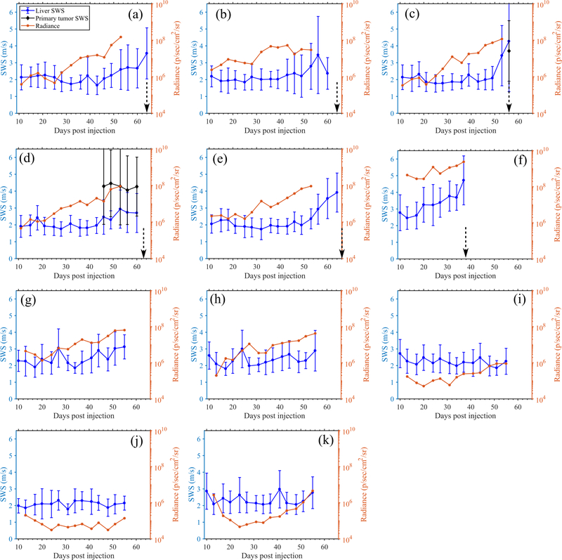
Fig. 12:
Quantitative longitudinal measurements obtained from the Gemcitabine treated mice using pSTL-SWEI and BLI. Results show the SWS of the liver, SWS of primary tumors ((c) and (d) only) and radiance measured in the abdomen. In SWS measurements, the errorbar indicate the spatial standard deviation of SWS. The dotted vertical arrows indicate the time of death. Mice corresponding to (i)-(k) were excluded from statistical analysis due to insufficient cell injection.

