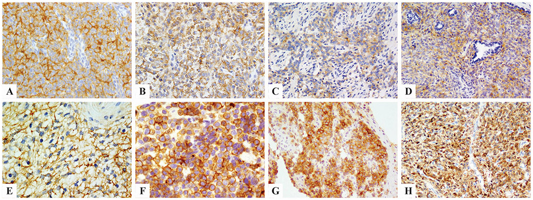
Figure 1. Immunohistochemical stains for pan-Trk.
showed diffuse moderate staining in YWHAE-rearranged sarcomas (A), variable staining in soft tissue round cell sarcoma with BCOR ITD (B), clear cell sarcoma of kidney with BCOR ITD (C), and BCOR-CCNB3 fused sarcomas (D). Ossifying fibromyxoid tumor harboring ZC3H7B-BCOR fusion showed diffuse but moderate cytoplasmic pan-Trk staining (E). One Ewing sarcoma with EWSR1-FLI1 fusion had diffuse membranous staining for both pan-Trk (F) and NTRK1 (G). A BCOR-negative, NAB2-STAT6 positive solitary fibrous tumor showed diffuse and strong pan-Trk staining (H).

