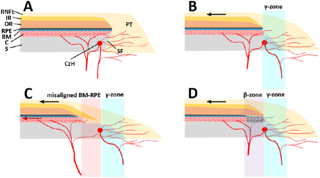
Figure 1.

Schematic illustrations of various parapapillary structures. (A) Nonmyopic eye without a γ-zone. (B) Myopic eye with a γ-zone but no nonjuxtapapillary MvD. Myopic axial elongation stretches the temporal parapapillary tissues (black arrow), resulting in the oblique SF presenting as a clinical γ-zone (light-blue region). Note that the SF within the γ-zone is abundant in microvessels supplying the prelaminar tissue (PT). (C) Myopic eye with a γ-zone and nonjuxtapapillary MvD associated with a misaligned BM–RPE complex. The BM-RPE complex is misaligned temporally with the adjacent inner retina (IR) and choroid (C), and further stretching potentially exceeds the elastic limit of the BM-RPE complex (red arrow). Note that the parapapillary endpoints of the BM-PRE complex, outer retina (OR), and C do not merge with the other tissues, whereas the end points of all retinal layers and C merge at the end point of the BM-RPE complex in other cases (A, B, D). The separation between the Circle of Zinn-Haller (CzH) and the end point of the BM-RPE complex is also notable. Although microvessels are still abundant in the deep layer (i.e., SF) of the γ-zone (light-blue region), they are scarce in the deep layer (i.e., nonjuxtapapillary sclera [S]) at the location of the misaligned BM-RPE (light-red region). (D) Myopic eye with a γ-zone and nonjuxtapapillary MvD associated with a β-zone. Although the microstructure is similar to that in the eye without nonjuxtapapillary MvD (B), choroidal perfusion is decreased in the β-zone (light-violet region) presenting as nonjuxtapapillary MvD. Note that microvessels are still abundant within the γ-zone (light-blue region). RNFL = retinal nerve fiber layer; IR = inner retina; OR = outer retina; RPE = retinal pigment epithelium; BM = Bruch's membrane; C = choroid; S = sclera; CzH = Circle of Zinn-Haller; SF = scleral flange; PT = prelaminar tissue.
