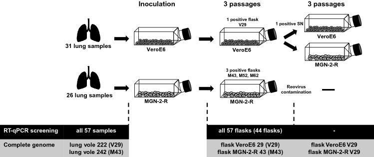
Fig. 1.
Schematic representation of the workflow. Bank voles were collected in forests within the district Osnabrück and a small piece of lung was taken by an incision in the chest area directly in the field. Lung tissue was meshed by grinding against a metal grid in a reaction tube containing 1 ml DMEM + 5% FCS and sterile filtered directly onto the cells. Cells were passaged three times until PUUV RT-qPCR screening. Supernatant of PUUV-positive flasks was taken and used for infection and further passaging in VeroE6 and MGN-2-R cells. Sequencing of complete genomes was done for PUUV RT-qPCR-positive passages and the corresponding original bank vole lung tissue. Isolates M52 and M62 were lost upon virus stock generation, presumably due to low viral load

