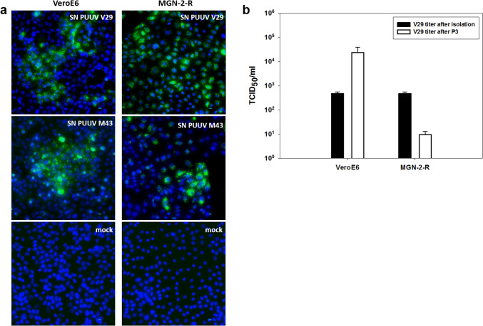
Fig. 2.
Infection studies of PUUV isolates in VeroE6 and MGN-2-R cells. a Immunofluorescence analysis of VeroE6 and MGN-2-R cells inoculated with supernatants (SN) of PUUV Osnabrück/V29, isolated on VeroE6 cells, or PUUV Osnabrück/M43, isolated on MGN-2-R cells. PUUV-inoculated and mock-infected cells were fixed 10 days post infection and stained with nucleocapsid protein-specific antibody 5E11 and a secondary anti-mouse Alexa fluor 488 conjugated antibody. Nuclei were stained with DAPI. b Determination of virus titers of PUUV Osnabrück/V29 isolate (TCID50/ml) after three passages (P3) in VeroE6 and MGN-2-R cells (white columns) in comparison to titers directly after isolation in VeroE6 cells (black columns). Titers were obtained by immunofluorescence staining of 96-well plates 10 days post inoculation

