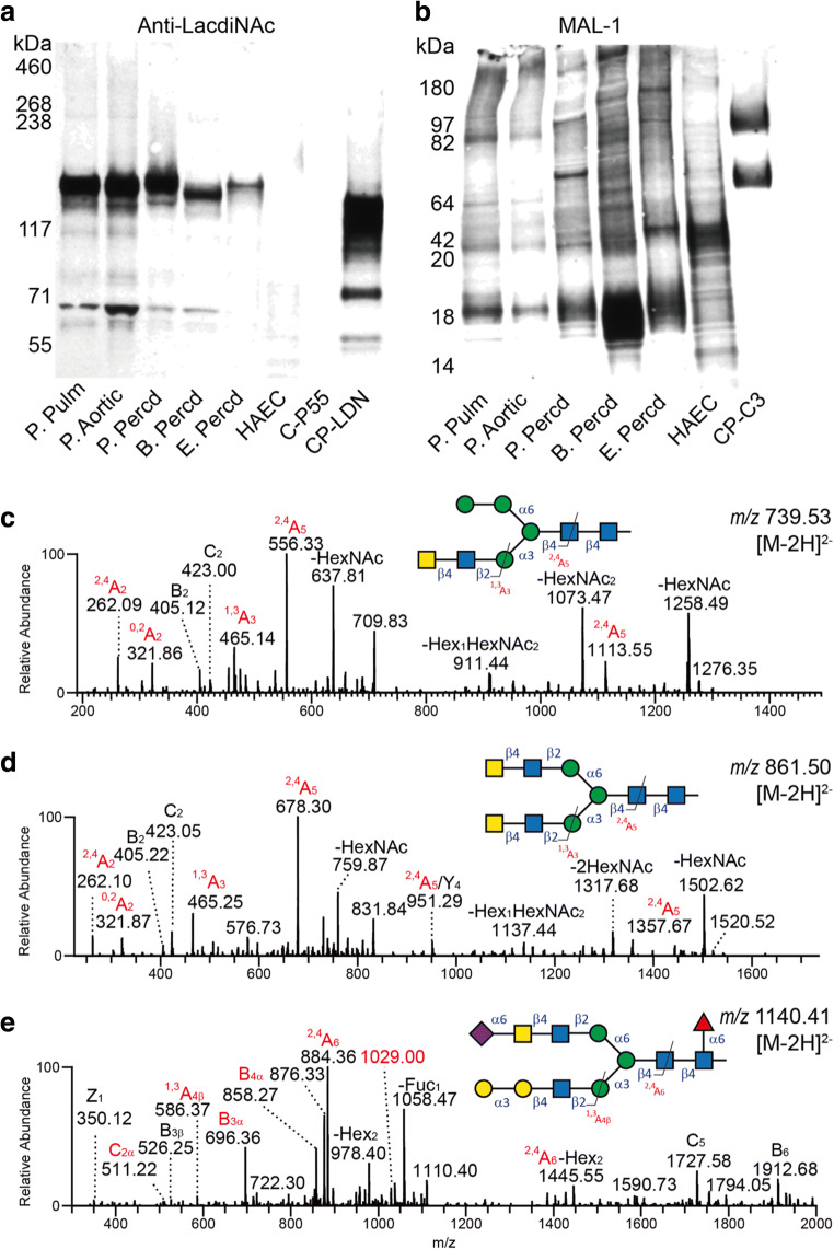
Fig. 4.
Western blot analysis of protein extracts from animal heart valves and pericardia using an anti-LacdiNAc antibody (a) and the MAL-1 lectin (b). A recombinant mucin-type fusion protein carrying terminal LacdiNAc determinants (CP-LDN) and purified from CHO-K1 cells transfected with plasmids encoding human B4GALNT3 and GCNT1 was used as a positive control. Positive control for MAL-1 staining was a mucin-type fusion protein carrying core 3 O-glycans extended with type 2 outer chains (CP-C3). A human aortic endothelial cell lysate was used as negative for LacdiNAc staining. MS/MS spectra of three LacdiNAc-containing N-glycans with masses corresponding to Hex4HexNAc4 ([M-2H]2- of m/z 739), Hex3HexNAc6 ([M-2H]2- of m/z 861), and Neu5Ac1Hex5HexNAc5deHex1 ([M-2H]2- of m/z 1140) are shown in c, d and e, respectively

