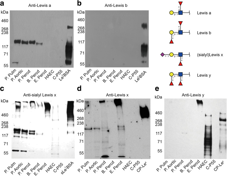
Fig. 5.
Western blot analysis of protein extracts from animal heart valves, pericardia and HAECs using anti-Lea (a), anti-Leb (b), anti-sialyl Lex (c), anti-Lex (d), and anti-Ley (e) antibodies. Positive control samples were Lea-, Leb- and sLex-BSA neoglycoconjugates. For Lex and Ley staining, recombinant mucin-type fusion proteins carrying Lex (CP-Lex) or Ley determinants (CP-Ley) were used as positive controls, while C-P55 was the negative control

