Figure 1.
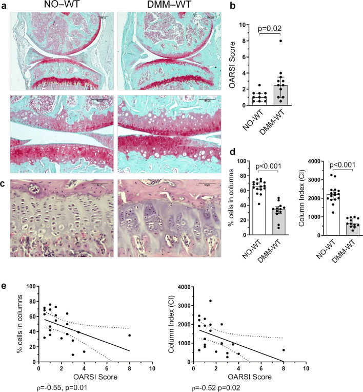
Disorganization of GP chondrocytes during experimental knee osteoarthritis (OA). Destabilization of medial meniscus (DMM) was induced in 12 weeks old mice, that were sacrificed 8 weeks after surgery. Post-traumatic damage accounted for the articular cartilage (AC) lesions, regarding proteoglycan loss, cartilage fibrillation and vertical clefts observed in the AC. (a) Representative mice joint sections stained with Safranin-O Fast Green at × 5 and × 20 magnification from non-operated wild type (NO-WT) and DMM-WT mice (bars = 250 and 100 µm respectively). (b) Histopathological OARSI Score represented as the sum of tibia and femur scores. (c) Representative Haematoxilin-Eosin stained tibia growth plate (GP) sections at × 40 magnification from NO-WT and DMM-WT mice (bars = 50 µm). (d) Percentage of cells in columns and column index (CI) in the GP in NO-WT and DMM-WT mice. Data are shown as median ± interquartile range (IQR). (e) Association between the percentage of cells in columns and OARSI score, and between CI and OARSI score in WT mice. NO-WT, n = 10–16; DMM-WT, n = 11–12.

