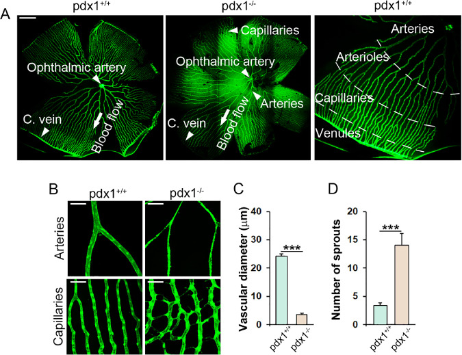
Figure 1.
Adult pdx1 mutants show vascular changes consistent with DR. (A) Retina flat mounts from middle-aged (18 months old) fli1a:EGFP transgenics, wild type (left) or pdx1−/− (center), and a close-up view of a wild type (right), with regions of the vascular plexus indicated. Arteries originating from the ophthalmic artery progressively branch to form a peripheral capillary plexus that drains into the circumferential vein (C. vein). Size bar indicates 500 µm. (B) Images of retinal vasculature in flat-mounted retinae from middle-aged adult (14–18 month old) pdx1+/+; fli1a:EGFP controls and pdx1−/−; fli1a:EGFP mutants focusing on central arteries (top) and peripheral capillaries (bottom). In mutants, arteries show narrowing, whereas capillaries have increased sprouts and branches. Size bars indicate 50 µm. (C) Quantitation of vessel diameter in 15 arteries from 4 individual fish per group, and (D) sprouting in 10 capillary regions from 3 individual fish per group, from samples as shown in B. ***P < 0.001.

