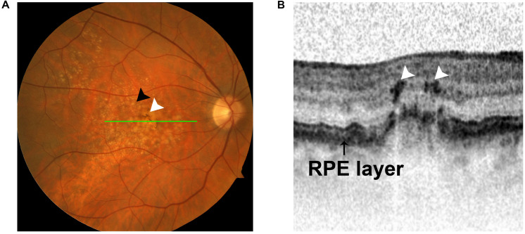
FIGURE 4.
Intraretinal hyperreflective foci (HRF) appear in OCT images in intermediate AMD. (A) Fundus photograph showing two hallmarks of intermediate AMD: sub-RPE deposits (black arrow) and pigment changes (white arrow). (B) OCT image demonstrating the presence of intraretinal HRF (white arrows) in intermediate AMD. The HRF correlate with pigment on funduscopic imaging (green line).

