
Fig. 2.
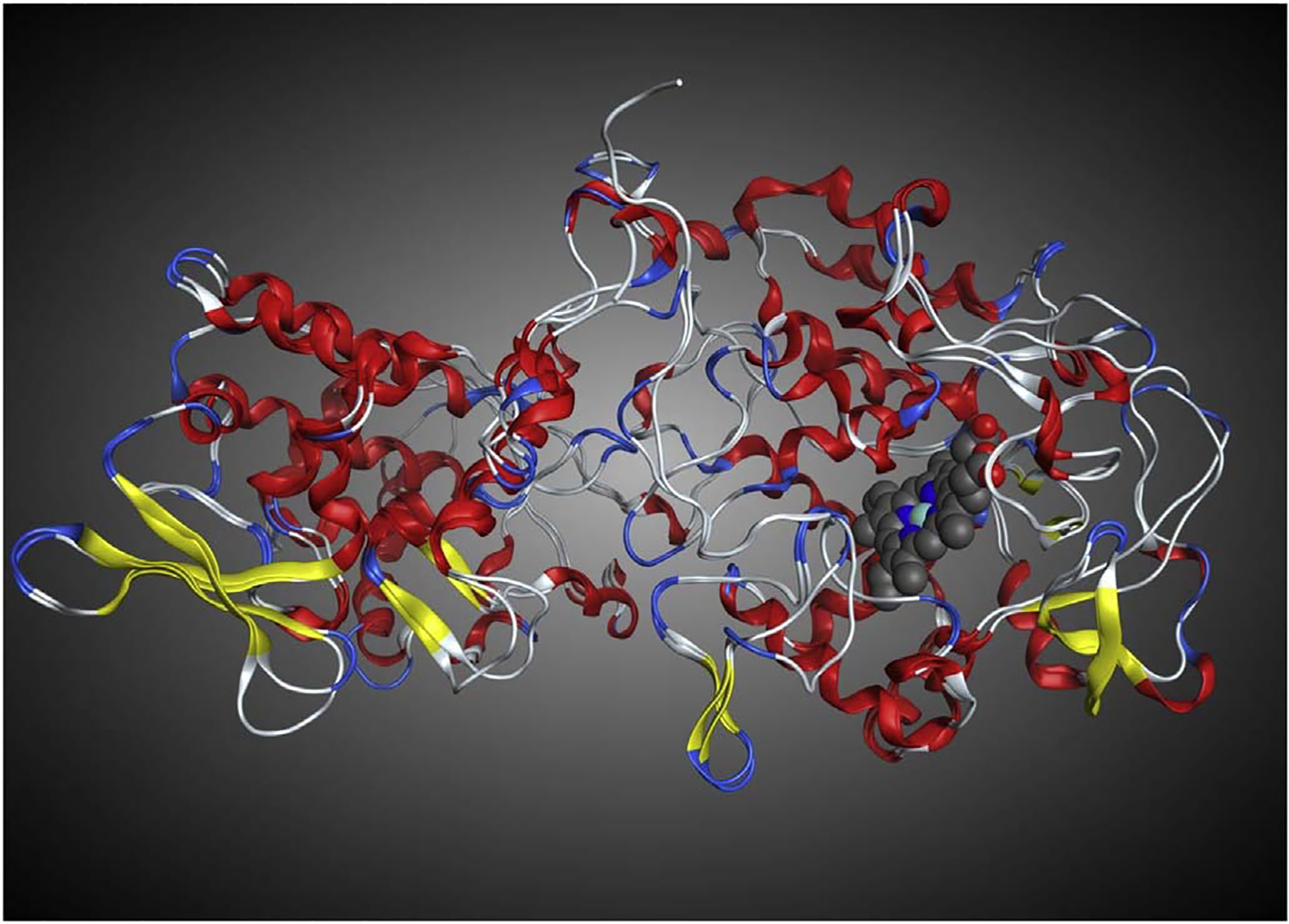
Structural superposition of the x-ray crystal structure of WT M. tuberculosis-KatG (2CCA.pdb) with WT Se-KatG (3WXO.pdb) illustrating the similar secondary and tertiary structures, overall 1.03 Å RMSD. The heme from 2CCA.pdb is shown in blue and grey. (For interpretation of the references to colour in this figure legend, the reader is referred to the web version of this article.)