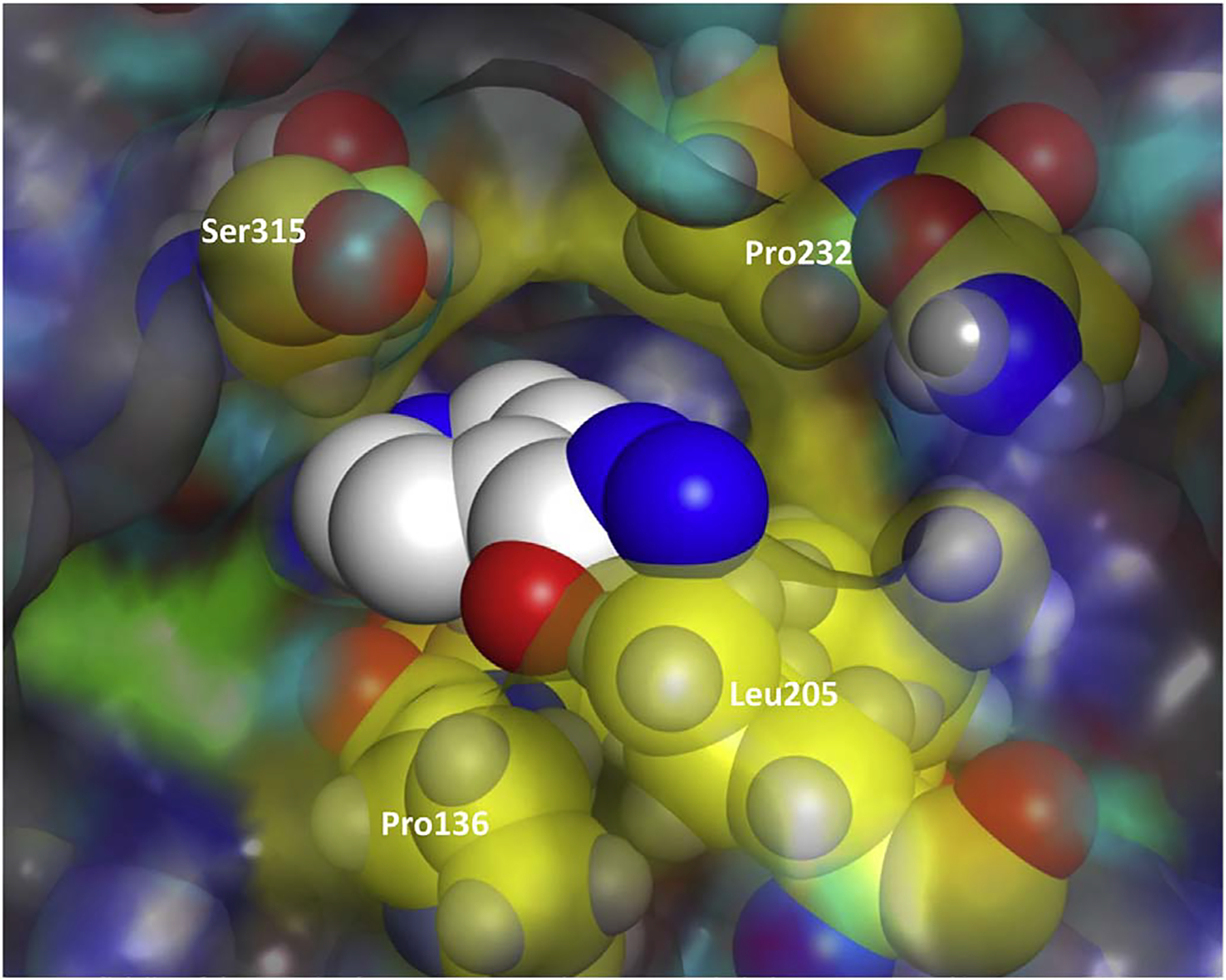
Fig. 4.

Channel 1 residues (in yellow) from WT M. tuberculosis-KatG (2CCA.pdb) superimposed on the x-ray structure of WT Se-KatG with bound INH (3WXO.pdb) in CPK coloring. (For interpretation of the references to colour in this figure legend, the reader is referred to the web version of this article.)
