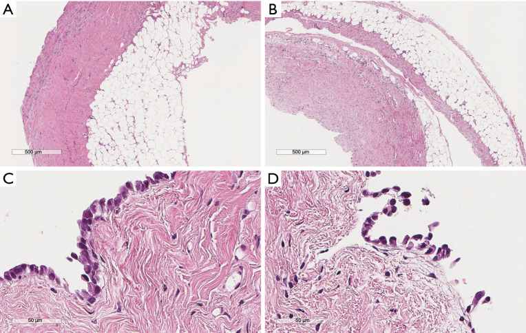
Figure 1.
The initial pleural biopsy of a patient with recurrent pleural effusion. Lower magnification images (A,B) show a slightly thickened fibrotic pleura and also the adequacy of the specimen which comprises the underlying adipose tissue. No tumor modules or any epithelial proliferation in noted in this examination. Higher magnification (C,D) shows a single mesothelial cell layer. Mesothelial cells show mild atypia. No invasive disease can be diagnosed in this specimen. Staining: hematoxylin, eosin, saffran.

