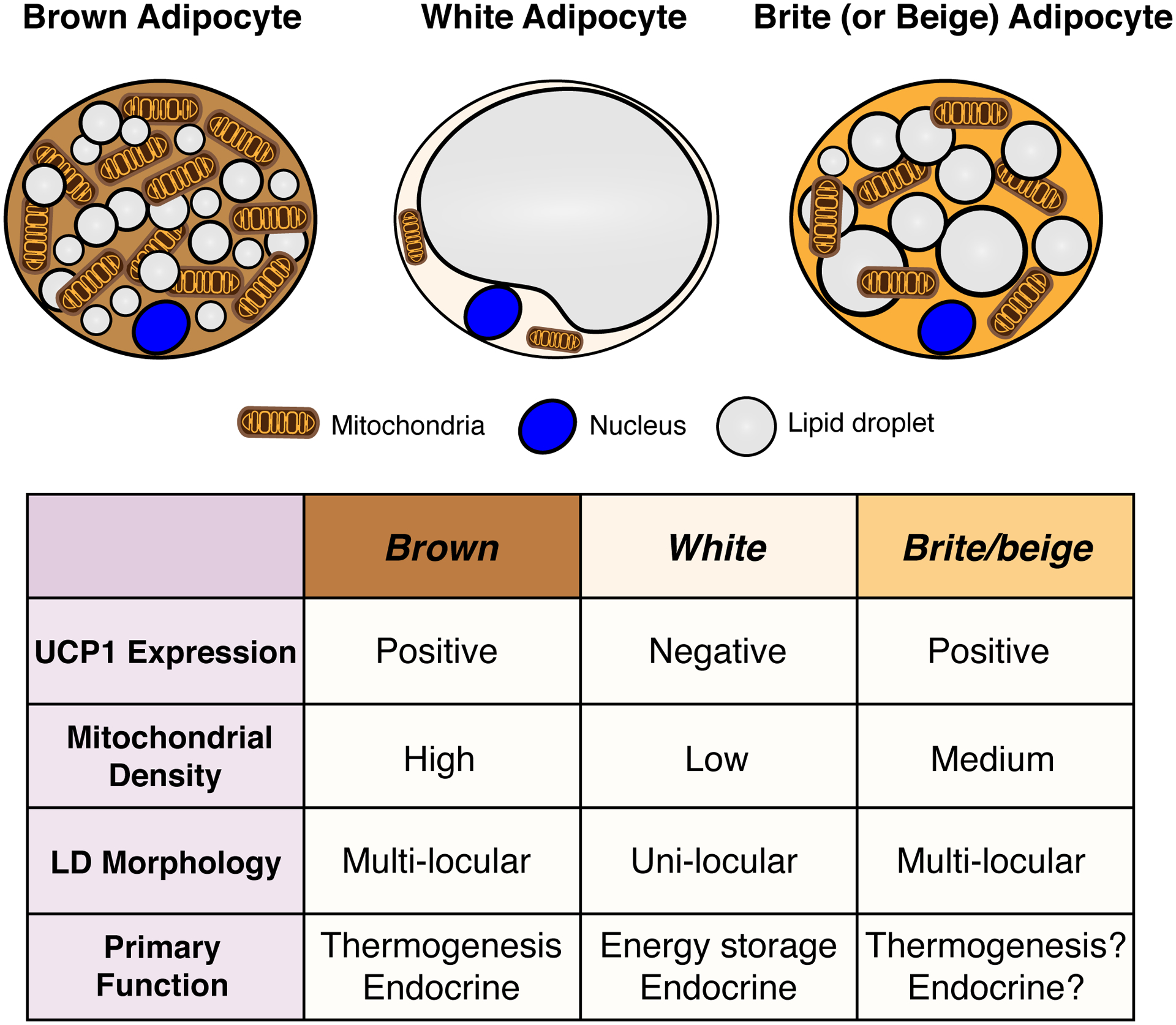
Figure 1. General characteristics of brown, white and brite/beige adipocytes.

A stimulated brown adipocyte (left) contains numerous small lipid droplets, many mitochondria, and expresses high levels of uncoupling protein 1 (UCP1), which is embedded in the inner mitochondrial membrane and required for thermogenesis. The color of brown fat reflects the high iron content of mitochondria. A white adipocyte (middle) in contrast contains a single large lipid droplet, fewer mitochondria, and does not express UCP1. A Brite/beige adipocyte (right) is characteristically intermediate between brown and white adipocyte, having multiple lipid droplets (though often larger than those seen in a brown adipocyte), more mitochondria than a white adipocyte, and it expresses UCP1.
