Figure 1.
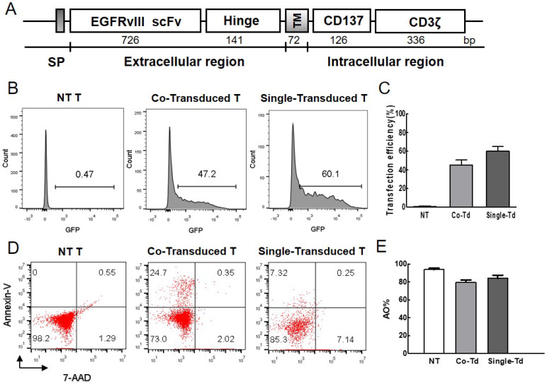
Evaluation of transfection efficiency and viability. (A) Structure of EGFRvIII CAR. It contained EGFRvIII scFv, the hinge and transmenbrane (TM) region of human CD8α, CD137 signaling domain, and human CD3ζ chain. IgG κ chain was used as signal peptide (SP). (B) The percentage of GFP-positive cells represented transfection efficiency of foreign gene at 24h after electroporation. Non-transduced cells were used as control. (C) Transfection efficiency was detected by flow cytometry (n=3). (D) Cell mortality post-electroporation was examined by flow cytometric analysis. Cells were stained with Annexin V-APC and 7-AAD dye, the fraction of Annexin+/7-AAD- dead cells was indicated. (E) AO/PI dual-fluorescence for live/dead staining was also used to quantify cell survival rate and detected by automated cytometer (n=3). Co-Transduced T (Co-Td): T cells was transfected with CAR-transposon and transpoase. Single-Transduced T (Single-Td): T cells was transfected with CAR-transposon. NT T: non-transduced T cells.

