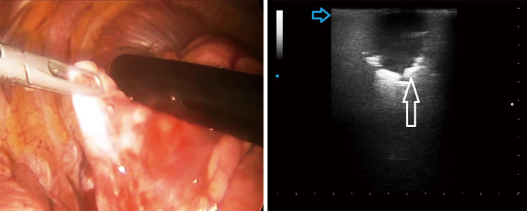
Figure 1.
Intra-operatory lung ultrasound during video-assisted thoracic surgery by a linear probe (12.5 MHZ) showing pulmonary hypoechoic nodule with irregular margin (white arrow) and increased thickness of the hyperechoic pleural line (blue arrow) with no artifacts below. Histologic diagnosis: adenocarcinoma and pulmonary fibrosis.

