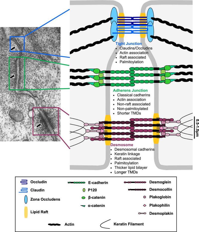
Figure 2: Intercellular junction structure, composition and key characteristics.
Depicted in the electron microscopy image of polarized rat intestinal mucosa [modified image from [135]], intercellular junctions are arranged characteristically with TJs (blue) at the most apical side followed by AJs (green) and then desmosomes (purple). TJs maintain polarity and regulate paracellular ion flow and are composed of the membrane proteins claudins and occludin which interact with intracellular zona occludens proteins and additional adaptors to link to the actin cytoskeleton. AJs mediate calcium-dependent adhesion by anchoring classical cadherin transmembrane proteins to the actin cytoskeleton through intracellular adaptor proteins. Desmosomes also mediate calcium-dependent adhesion but can attain a stronger, calcium-independent state, allowing tissues to resist mechanical stress. Desmosomal cadherin transmembrane proteins are anchored to the intermediate filaments through intracellular adaptor proteins to mechanically couple adjacent cells.

