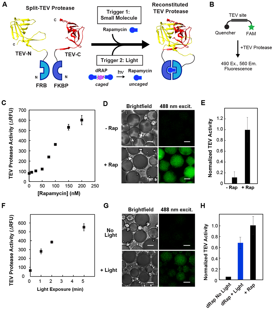
Fig. 3. Activation of split TEV protease in cell-like compartments using small molecule and light-inducible dimerization.

(A) Schematic of photocaged rapamycin, dRap, and split TEV fragments fused to FRB and FKBP. (B) Schematic of TEV activity assay: upon substrate cleavage, a FAM fluorophore is released from quencher. (C) Dose-dependence of rapamycin mediated TEV reconstitution. Assay uses 125 nM of split TEV proteins and varying concentrations of rapamycin. (D-E) Chemically induced reconstitution of TEV activity inside celllike compartments. Equimolar concentration of rapamycin promotes TEV protease activity; there is low background activity in the absence of rapamycin. (F) Optical uncaging of dRap at various exposure times, promotes TEV reconstitution in a plate reader assay. 125 nM split TEV and 73 nM dRap. (G-H) Temporal triggering of TEV activation within cell-like compartments using light. 10 min exposure to 365 nm UV light to uncage dRap within emulsions. Minimal background activity in non-illuminated samples. For 3D-E and 3G-H, 500 nM split TEV proteins with equimolar rapamycin or equivalent dRap. For 3G-H, activity from a control without dimerizer was subtracted from conditions with dimerizer present. Scale bar: 5 μm.
