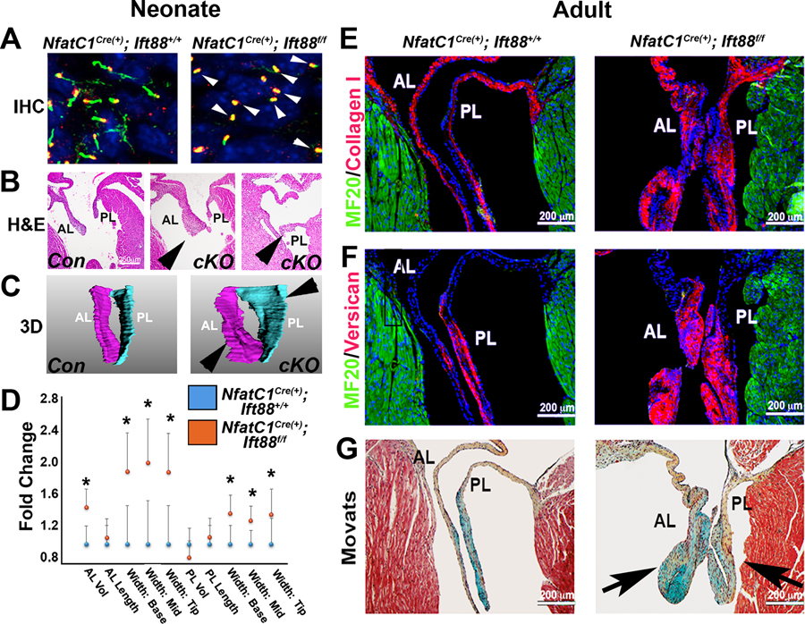
Fig. 2. Loss of Ift88 impairs ciliogenesis during development and results in myxomatous mitral valve disease.
(A) IHC for acetylated tubulin (green) and basal bodies (red) at neonatal day 0 in conditional knockout for Ift88 [NfatC1Cre(+);Ift88f/f] and control mice shows axoneme structures (loss indicated by arrowheads). (B) Hematoxylin and eosin (H&E) staining for Ift88 conditional knockout (cKO) and control mice at P0. (C) 3D reconstructions of the mitral valve anterior and posterior leaflets (valve thickening indicated by arrowheads). (D) Quantification of valve 3D reconstructions of control [ NfatC1Cre(+);Ift88+/+] and Ift88 conditional knockout [ NfatC1Cre(+);Ift88f/f] mitral leaflets. *P < 0.03; n = 6 for conditional knockout and n = 4 for control. Dots represent means, and error bars are SD of the mean. (E) IHC for collagen (red), myocardium (green), and nuclei (blue) on adult Ift88-deficient mitral leaflets and controls. (F) IHC for versican (red), myocardium (green), and nuclei (blue) on adult Ift88-deficient mitral leaflets and controls. (G) Movats histological stain on adult Ift88-deficient mitral leaflets and controls [arrows indicate increased proteoglycans (blue) and enlarged valve leaflets] compared to controls. Proteoglycan, blue; collagen, yellow; myocardium, red. n = 4 per genotype.

