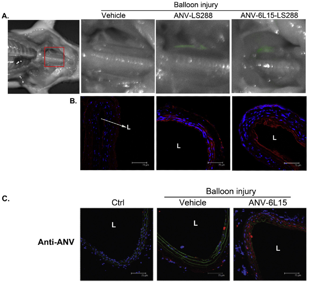
Fig. 1.

Binding of ANV-LS288 and ANV-6L15-LS288 to the balloon-injured carotid artery. A. Near infrared (near-IR) fluorescence imaging was carried out 8 h after balloon injury of the left external carotid artery and i.v. bolus injection of vehicle (PBS), ANV-LS288 (50 μg/kg), or ANV-6L15-LS288 (50 μg/kg). The balloon injured, but not the uninjured, carotid arteries showed near-IR (pseudo-color: green) fluorescence due to the binding of ANV-LS288 or ANV-6L15-LS288. B. Immunohistological staining of the injured carotid artery sections using anti-ANV antibody, and Cy3-conjugated secondary antibodies (red), and DAPI nucleus stain (blue). The anti-ANV antibody detected the bound ANV-LS288 or ANV-6L16-LS288 on the injured arterial wall. L: lumen. C. Immunohistological detection of ANV-6L15 bound to the balloon-injured carotid artery 24 h after i.v. bolus injection. The left external carotid artery of rats was injured by balloon angioplasty and treated with PBS vehicle or a bolus of ANV-6L15 (50 μg/kg). After 24 h, the rats were sacrificed and the uninjured and balloon-injured carotid artery sections were immuno-stained with anti-ANV antibody and Cy3-conjugated second antibody (red). The uninjured carotid artery (Ctrl) barely showed red color stain; the PBS-treated balloon-injured carotid artery (Vehicle) lightly stained red; and the ANV-6L15-treated balloon-injured carotid artery (ANV-6L15) stained red intensely. L: lumen.
