
Figure 3.
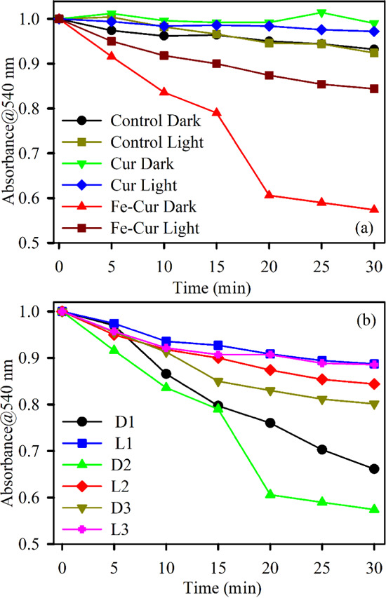
Absorption kinetics of DPPH degradation (monitored at 540 nm) (a) in the absence of light using samples Cur (green) and Fe–Cur (red) and in the presence of green light with Cur (blue) and Fe–Cur (brown) and (b) in the absence (D1: 0.07 OD, D2: 0.10, and D3: 0.15 OD) and presence (L1: 0.07 OD, L2: 0.10 OD, and L3: 0.15 OD) of light using samples Fe–Cur with variable concentrations.