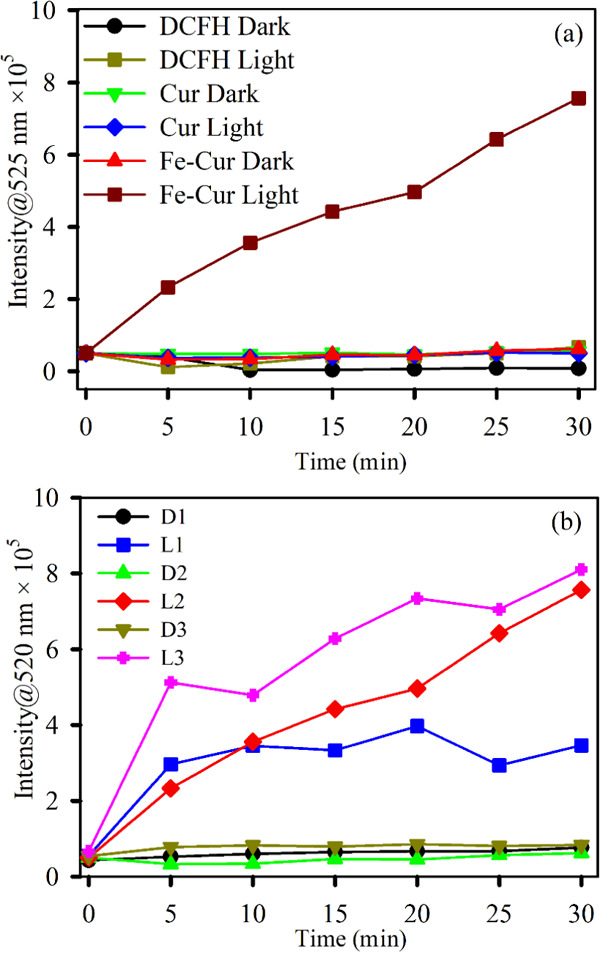
Figure 4.

DCFH oxidation (monitored at 525 nm) with time (a) in the absence of light with samples Cur (green) and Fe–Cur (red) and in the presence of light with Cur (blue) and Fe–Cur (brown) and (b) in the absence (D1: 0.07 OD, D2: 0.10 OD, and D3: 0.15 OD) and presence (L1: 0.07 OD, L2: 0.10 OD, and L3: 0.15 OD) of light using samples Fe–Cur with variable concentrations. The excitation wavelength was 488 nm.
