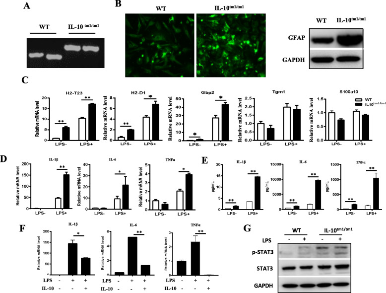
Fig. 2.
Astrocytes from IL-10tm1/tm1 mice were prone to the A1 phenotype. a Genotyping of WT and homozygous IL-10tm1/tm1 mice. b Representative immunofluorescent staining (right) and representative western blots for GFAP in astrocytes from WT and IL-10tm1/tm1 mice. Representative graph showing relative mRNA levels of indicated transcripts (c) and pro-inflammatory factors (d) in primary cultured astrocytes isolated from WT and IL-10tm1/tm1 mice stimulated with or without 100 ng/mL LPS for 24 h. e Protein levels of the indicated cytokines in the supernatants of primary cultured astrocytes isolated from WT and IL-10tm1/tm1 mice which stimulated with or without 100 ng/mL LPS for 24 h. f Representative graph showing relative mRNA levels of indicated pro-inflammatory cytokines in astrocytes isolated from IL-10tm1/tm1 mice treated with the indicated factors. Data shown are means ± SD of two independent experiments. g Representative western blots showing the expression of indicated proteins in astrocytes from WT and IL-10tm1/tm1 mice treated with or without LPS. *p < 0.05 and **p < 0.01

