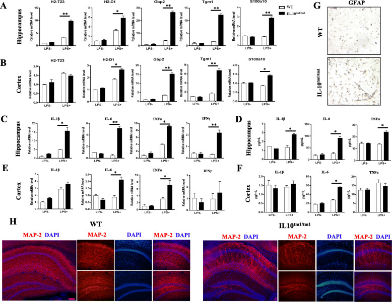
Fig. 4.
Higher expression of A1-specific transcripts and pro-inflammatory factors and severe neuron death in IL-10tm1/tm1 mice. Representative graph showing relative mRNA levels of indicated transcripts (a, b) and pro-inflammatory factors (c, e) in the hippocampus (a, c) and cortex (b, e) from WT and IL-10tm1/tm1 mice with i.p. injection with or without LPS. Protein levels of the indicated cytokines in the hippocampus (d) and cortex (f) from WT and IL-10tm1/tm1 mice subject to i.p. injection with or without LPS. g Representative immunohistochemistry staining for GFAP in WT and IL-10tm1/tm1 mice. f Representative immunofluorescent staining for MAP2 in the hippocampus of WT and IL-10tm1/tm1 mice. *p < 0.05 and **p < 0.01

