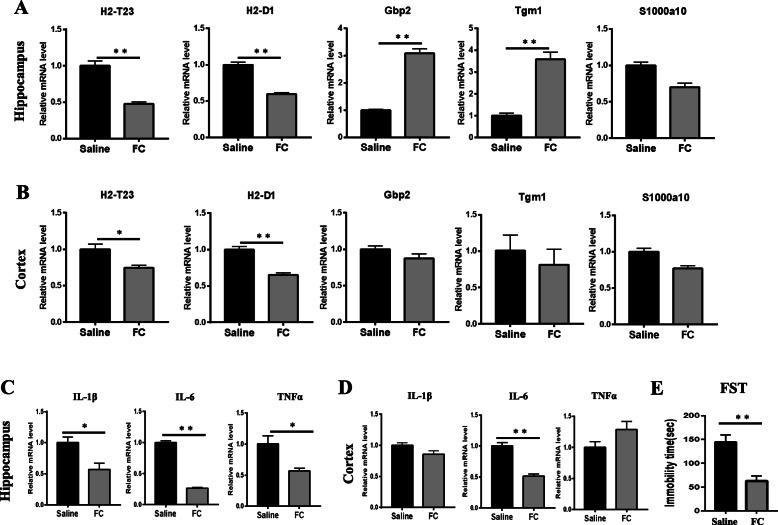
Fig. 7.
FC treatment inhibited A1 astrocyte activation and ameliorated LPS-induced behavior deficits Mice were injected i.c.v with normal saline or FC solution (1 nmol per mouse), and 6 h later, mice also received either an i.p. injection of saline or LPS (0.83 mg/kg). The FST was performed 24 h following administration of LPS. Immediately following behavioral tests, mice were killed and perfused with ice-cold phosphate-buffered saline, and the hippocampus (a, c) and cortex (b, d) were rapidly collected and measured by real-time PCR for transcription factors (a, b) and inflammatory factors (c, d). The duration of immobility during the FST (e) was recorded 24 h following administration of LPS. Data were expressed as the mean ± SD. *p < 0.05 and **p < 0.01

