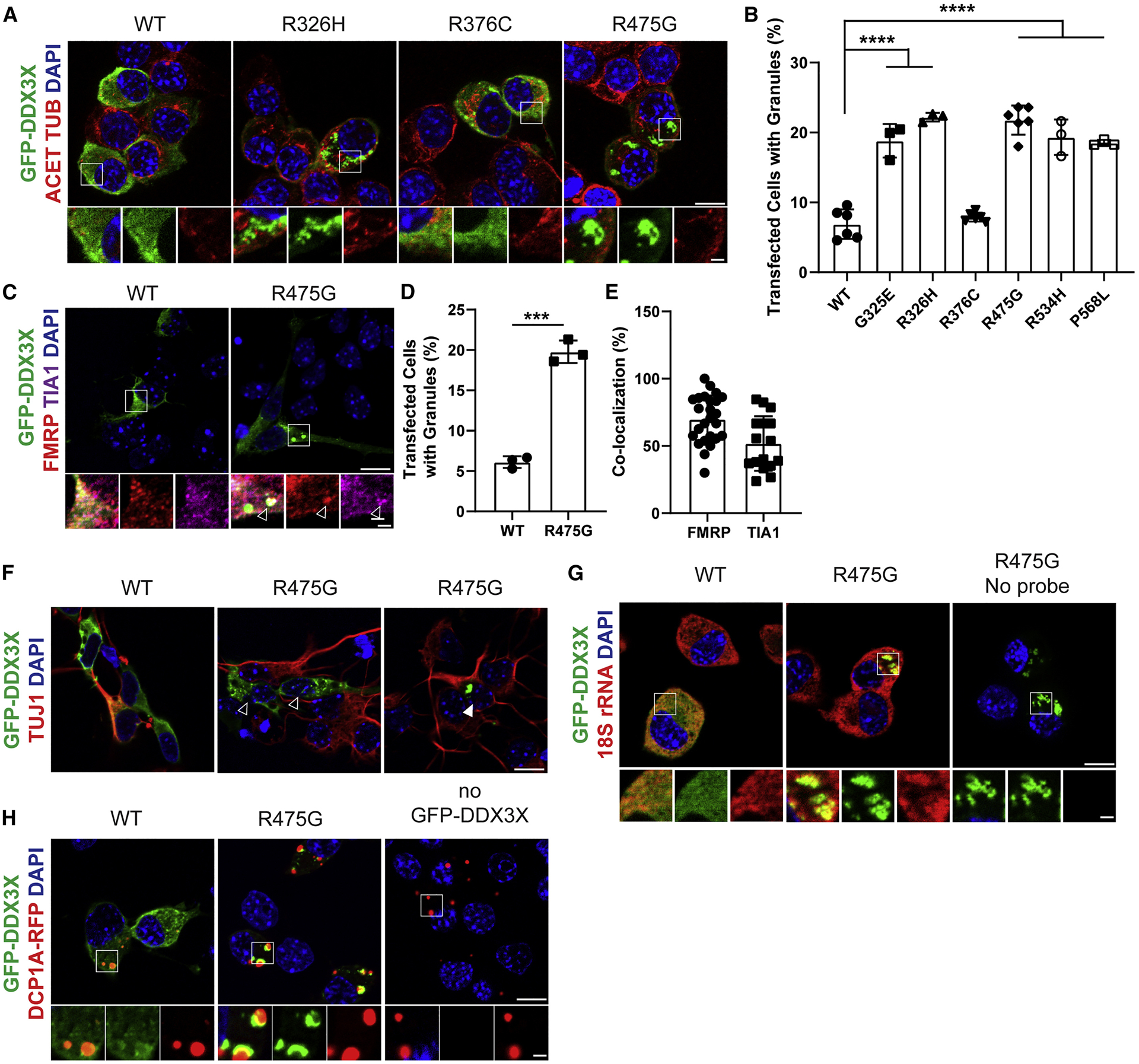
Figure 6. DDX3X missense mutations induce ectopic RNP granules in neural progenitors.

A, Images of N2A cells transfected for 24 hrs with WT or mutant DDX3X-GFP and immunostained for GFP (green), Acetylated-TUBULIN (red), and DAPI (blue). Below, high magnification images from boxed regions. B, Quantification of percentage of N2A cells containing WT or mutant DDX3X-GFP granules. C, Primary cortical cells transfected for 24 hrs with WT or R475G DDX3X-GFP (green) and co-stained for FMRP (red) and TIA1 (magenta), with granule co-localization granules (arrowhead). D, Quantification of percentage of primary cortical cells containing DDX3X-GFP granules. E, Quantification of DDX3X-granules co-localized with RNA-binding proteins TIA1 or FMRP. F, Primary cortical cells expressing either WT or R475G DDX3X-GFP (green) and stained with TUJ1 (red). Both TUJ1- progenitors (empty arrowhead) and TUJ1+ neurons (filled arrowhead) contain DDX3X granules. G, N2A cells transfected with DDX3X-GFP (green) and probed for 18S rRNA with smFISH probes (red). H, N2A cells transfected with DDX3X-GFP (green) and DCP1A-RFP (red) to mark P-bodies. Scale bars, 10 μm (low magnification) and 2 μm (high magnification) (A, C, F, G, H). Error bars=SD.
