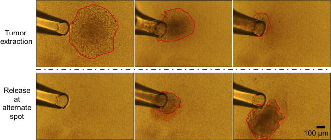
Figure 5.
A tumor on top of the collagen I island can be easily extracted. Top: Successive images of a tumor being extracted from a collagen island, after 20–30 min treatment with collagenase, using a glass tip of ~ 150 µm diameter; Bottom: Successive images of the same tumor being released at an alternate location (e.g., for applications such as dissociation and re-seeding of selected single cells from the tumor). The red outline delineates the visible portion of the tumor during different steps of the process. (see video in Supplementary movie).

