Figure 5.
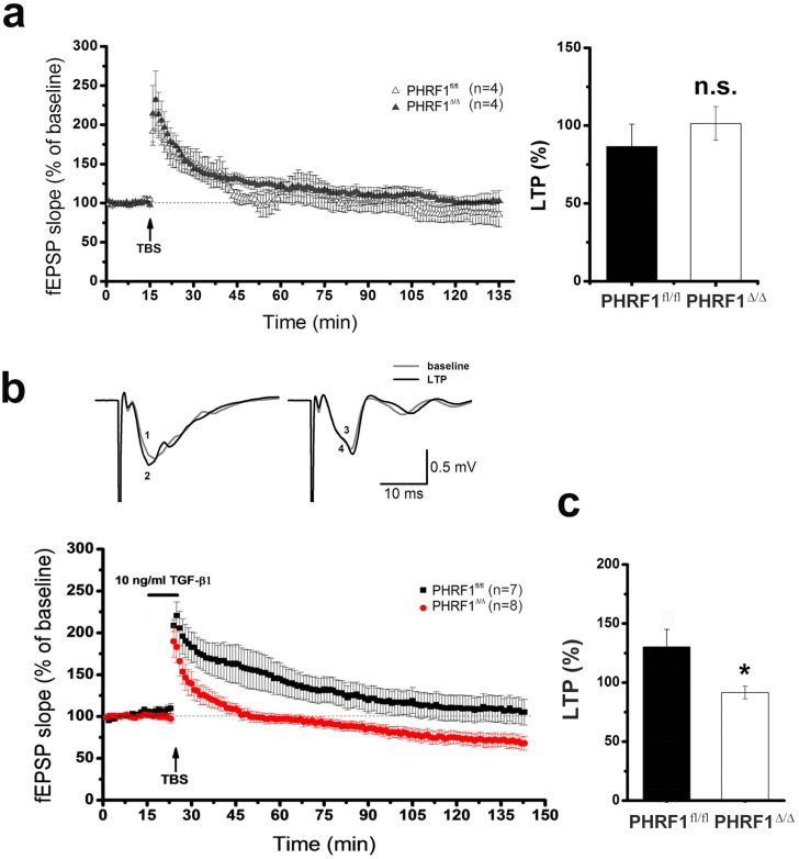
Theta burst stimulation (TBS) induced synaptic changes in the hippocampal slices. The extracellular field-EPSP (fEPSP) was recorded in hippocampal slices following the stimulation of the Schaffer collateral pathway. (a) An arrow indicates the start of theta burst stimulation (TBS). Note that there is no statistical difference (n.s.) between genotypes. (b) The hippocampal slices were incubated with TGF-β1 (10 ng/ml) for 15 min before the application of TBS (arrow). The black and red dots represent averaged points of fEPSP slope from PHRF1fl/fl and PHRF1Δ/Δ mice, respectively (n = 7 slices from 4 PHRF1fl/fl mice; n = 8 slices from 4 PHRF1Δ/Δ mice). Upper panel shows the representative tracings before (grey) and after (black) TBS of PHRF1fl/fl and PHRF1Δ/Δ mice. (c) At 60 min after TBS, the mean fEPSP values were analyzed by one-way ANOVA. Data are mean ± SEM. *p < 0.05.

