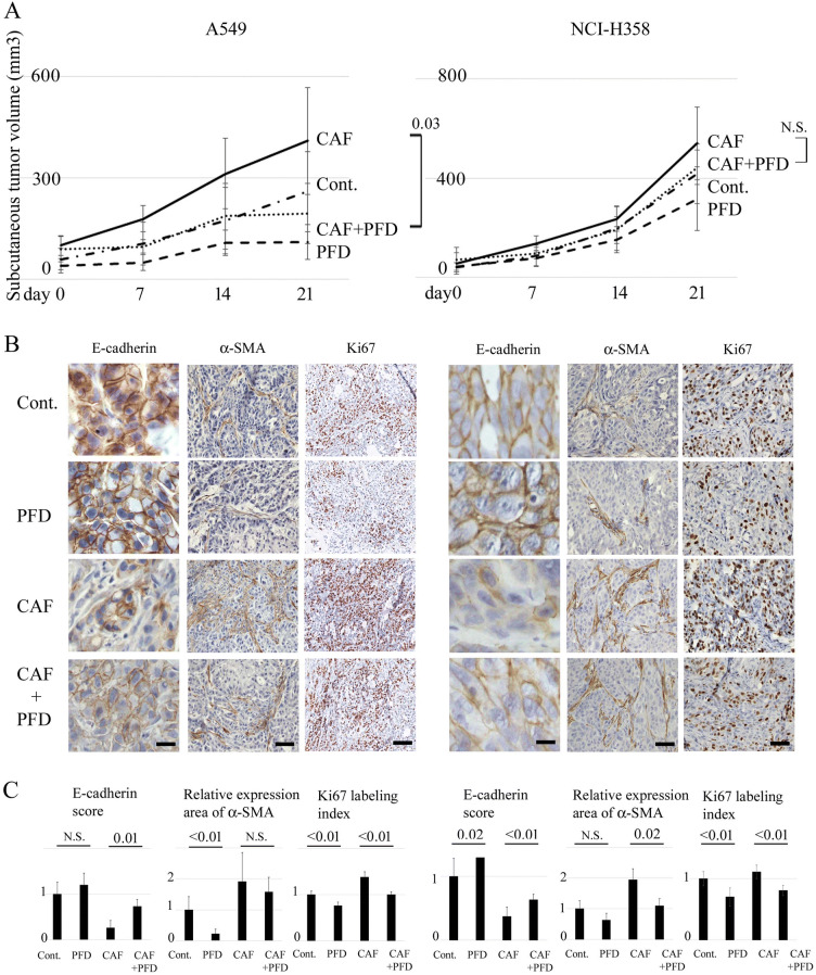
Figure 3.
Effects of pirfenidone on subcutaneous tumor formation of NSCLC cells and CAFs in vivo. (A) Tumor volumes (mean ± SD) after subcutaneous injection of A549 or NCI-H358 cells with or without CAFs into nude mice are plotted, and significance tested with repeated measures ANOVA. Mice are treated intraperitoneally with 200 mg/kg pirfenidone (PFD) or sterile water daily for 3 weeks. Each group contains six mice. The chain shows the tumor volume of control mice (Cont.): NSCLC cells only are transplanted without PFD treatment. Broken line (PFD): NSCLC cells only are transplanted with PFD treatment. Solid line (CAF): both NSCLC cells and CAFs are transplanted without PFD treatment. Dotted line (CAF + PFD): both NSCLC cells and CAFs are transplanted with PFD treatment. (B) Panels show representative findings of histological evaluation for E-cadherin, α-SMA, and Ki67 of primary tumors obtained from mice. Scale bar: 80 µm for E-cadherin, 200 µm for α-SMA, and 400 µm for Ki67. (C) Panels show the scored expression of E-cadherin, the relative area of α-SMA expression, and the Ki67 labeling index as the mean ± SD. Significance was tested with the Mann–Whitney U test.

