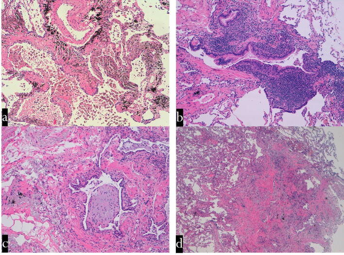
Figure 3.
Transbronchial cryobiopsy. Histological findings (haematoxylin–eosin staining: mid power). (a) Respiratory bronchiolitis: a smoker's macrophages in the lumen of the respiratory bronchiole and surrounding alveoli. The wall of the respiratory bronchiole is thickened by collagen deposition. (b) Follicular bronchiolitis: lymphoid follicles are evident in the wall of a terminal bronchiole. The lumen of this airway is partly occluded. (c) ILD with prominent bronchiolar involvement: The lumen of a terminal bronchiole is almost completely occluded by a polyp made up mainly of loose connective tissue (bronchiolitis in HP). (d) Constrictive bronchiolitis: a terminal bronchiole is completely substituted by a scar. *HPE histopathological examination, GL ILD granulomatous lymphocytic interstitial lung disease, CVID common variable immunodeficiency, GGO ground-glass opacity, NSIP nonspecific interstitial pneumonia, ILD interstitial lung disease, ANCA—anti-neutrophilic cytoplasmic antibody, OP organizing pneumonia, COPD chronic obstructive pulmonary disease, MAC—mycobacterium avium complex, HP hypersensitivity pneumonitis, DIPNECH diffuse idiopathic pulmonary neuroendocrine cell hyperplasia, CT computed tomography, DIP desquamative interstitial pneumonia, ILD smoking-related interstitial lung disease.

